


Notification

THANK YOU FOR MAKING AN APPOINTMENT AT AIH.
Would you like to pre-register?
Please fill in the information below
Urgent
DON'T BE SUBJECTIVE WITH BENIGN BREAST LUMPS, ACTIVELY SCREEN EARLY AND TREAT PROMPTLY AT AIH

Six months ago, patient S.K (22 years old, Cambodian nationality) discovered a small, painless lump in her left breast. However, recently the lump grew rapidly and caused pain, making the patient anxious, so she decided to visit American International Hospital (AIH) for a specialized examination.
At AIH, during the examination, Dr. Mai Thanh Cuc, a Specialist Level II in Oncology, noted a left breast mass approximately 3.5cm in size, multi-lobulated, and several masses measuring 6-8mm in the right breast. Combined with ultrasound findings, the doctor concluded bilateral multiple breast masses, with the left breast mass measuring 32 x 30 x 20mm, classified as BI-RADS 3. Subsequently, a fine needle aspiration (FNA) was performed, suggesting a benign fibroadenoma.
According to Dr. Mai Thanh Cuc, Level II Specialist, although this case was diagnosed as benign, because the tumor grew rapidly, was larger than 2cm, and the patient wanted the tumor removed due to anxiety and to prevent it from growing quickly and causing compression. Therefore, the patient was indicated for lesion removal using VAB (Vacuum-Assisted Biopsy) - a technique that suctions the lesion with a special needle assisted by a vacuum machine to remove the lesion without the need for a large incision, causing less pain, minimal damage to surrounding tissue, less scarring, and high cosmetic effectiveness.
The VAB technique, performed safely and conveniently by the team of doctors at the Oncology - General Surgery Center at AIH, pathology results showing it was a benign breast fibroadenoma. Thanks to the application of the minimally invasive VAB technique, the patient's health stabilized quickly, and they returned to normal activities within a short period of 24-48 hours.

Don't be subjective with benign breast lumps
Many women, upon discovering a breast lump, often feel reassured when their doctor says it's “benign”. However, subjectivity or neglecting follow-up can lead to unnecessary risks. In reality, some complex benign lumps can still increase the risk of breast cancer, or mask malignant lesions if not accurately and periodically checked.
Why should benign breast lumps not be underestimated?
Some complex benign tumors still slightly increase the risk of breast cancer
Lesions such as complex fibroadenomas and atypical hyperplasia can slightly increase the risk of breast cancer. Screening and monitoring help detect any abnormal changes early.
Benign tumors can obscure a malignant lesion, though rarely (only 0.0125%), such as malignant transformation within the epithelial component of a benign fibroadenoma, which can develop into lobular carcinoma in situ LCIS or DCIS.
If not thoroughly evaluated by ultrasound, mammography, or biopsy when necessary, cancer can be missed.
Timely treatment helps limit complications
If benign tumors grow rapidly or become large, they can cause pain, compression, breast deformation, or psychological impact. Doctors may prescribe monitoring, medication, or surgical intervention depending on the case, helping patients feel at ease and avoid unnecessary complications.
Signs that require immediate medical examination:
Appearance of a new lump in the breast
Rapidly growing or persistently painful lump
Nipple discharge, especially bloody discharge
Dimpling, wrinkling, redness, or orange peel-like skin on the breast
Retracted or shape-changing nipple
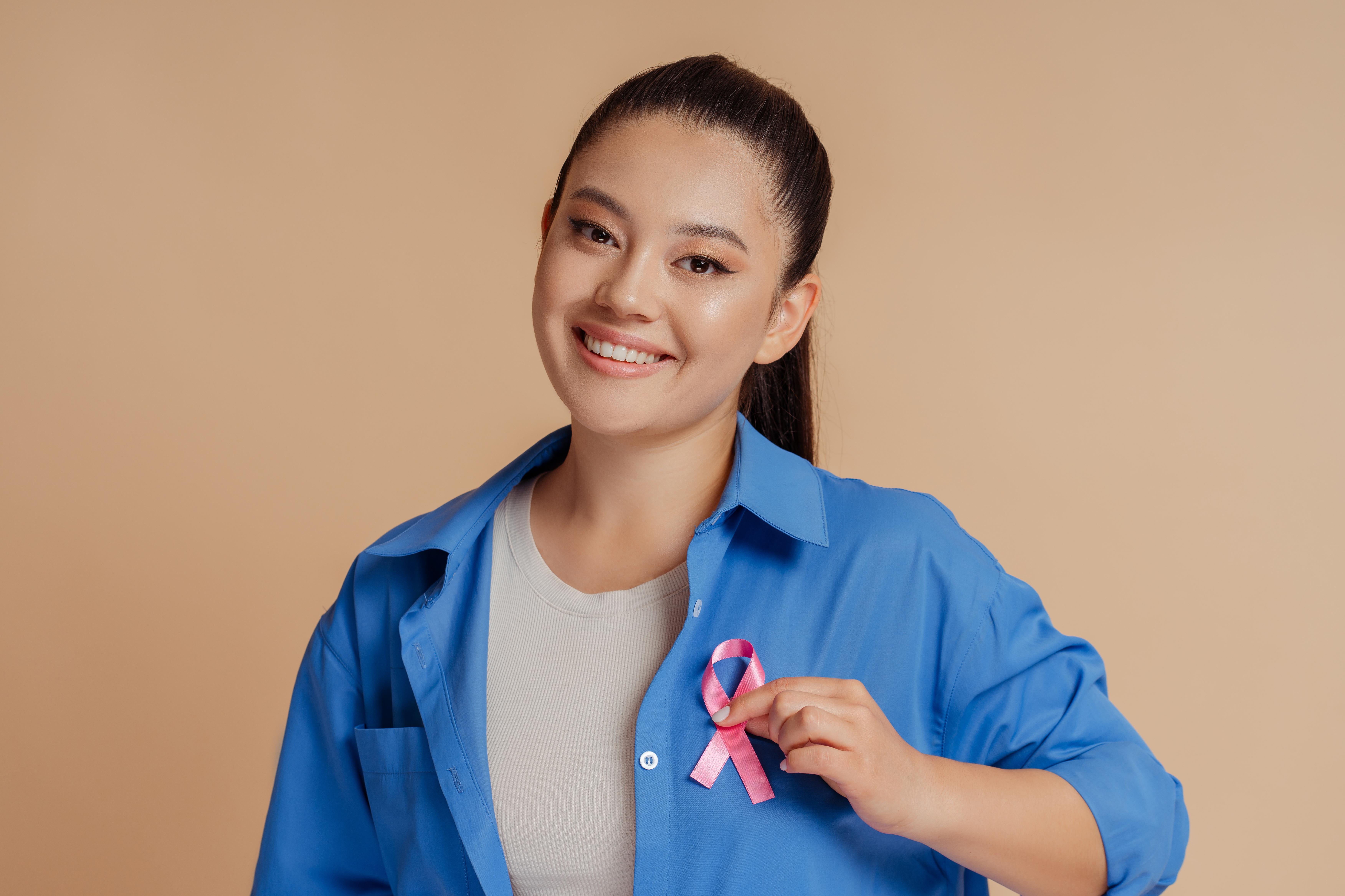
Screening – An unmissable importance
Women aged 20–39: Perform monthly breast self-examinations; undergo ultrasound if abnormal signs are present.
From 40 years old and above: Regular annual mammograms are recommended, or as advised by a doctor.
High-risk individuals (hereditary, family history, histological abnormalities...) need earlier and more frequent screening.
When is surgery necessary?
According to Dr. Mai Thanh Cuc, Level II Specialist, surgery will be indicated when the tumor is large (usually over 2-3 cm), grows rapidly in a short period; or causes pain or deformation of breast tissue, or when diagnostic imaging cannot completely rule out malignancy; or at the patient's request to remove it due to cancer concerns.
Interventional methods: VAB can be used for masses <2-3cm, typically benign, to avoid open surgery; or minor surgery if the mass is large, difficult to access, multiple tumors.
In the first 1-2 weeks, the patient will have a follow-up appointment to check the incision, infection, and the breast tissue. After 2-3 months, breast tissue and scar evaluation will be performed, followed by an ultrasound check after 6-12 months to detect early recurrence. From this point forward, the patient will continue annual periodic check-ups as guided by their doctor.
BREAST DISEASE SCREENING AND TREATMENT AT AIH – ACCURATE, SAFE, AND COMPREHENSIVE
At American International Hospital (AIH), the process of screening, diagnosing, and treating breast diseases is performed accurately and individualized for each patient, based on a modern imaging equipment system and close coordination between specialties, helping to detect lesions early, guide effective treatment, and ensure maximum safety.
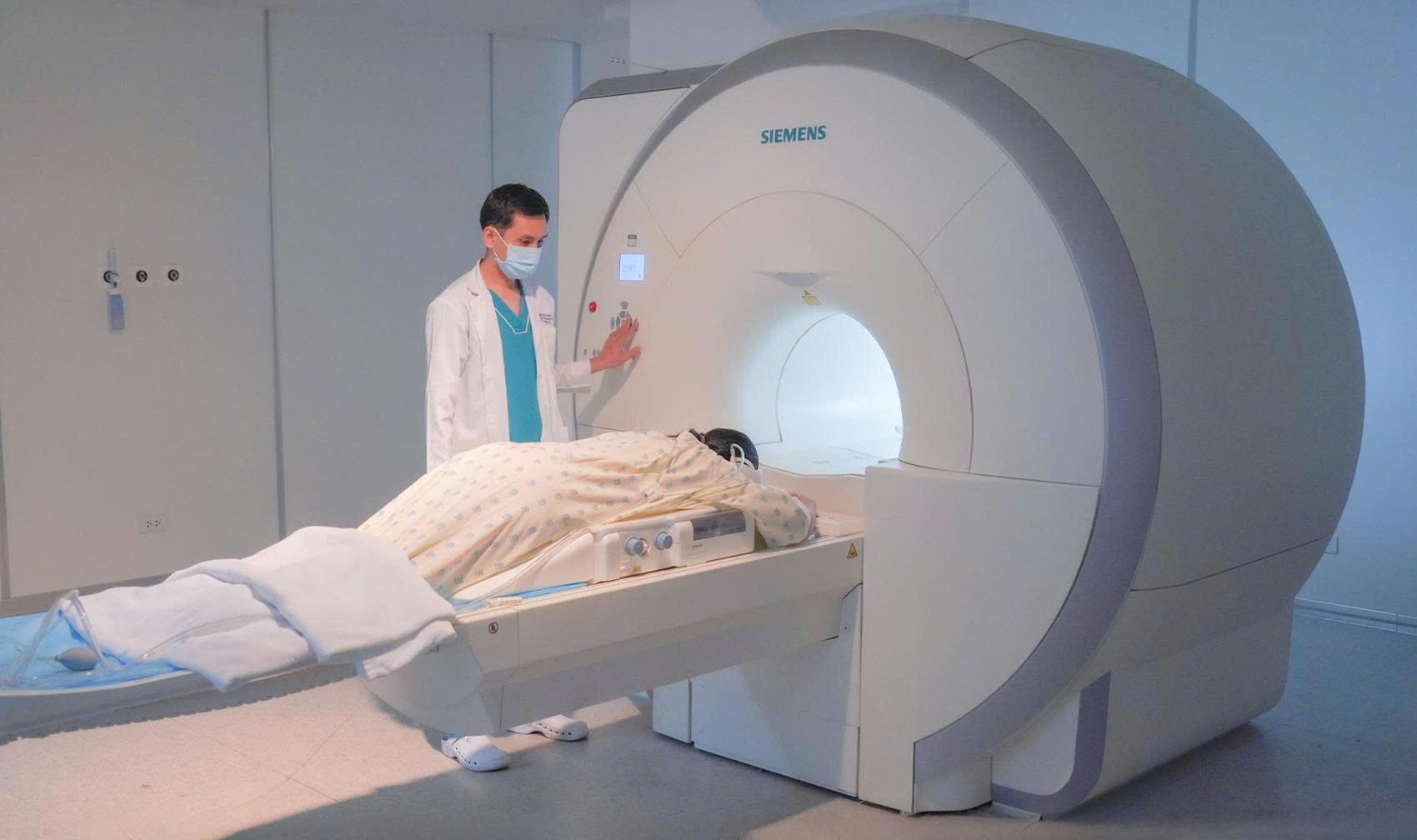
Advanced diagnostic technology: Specialized breast MRI application with dedicated Siemens signal receivers, aiding in accurate assessment of breast tissue lesions.
Experienced specialist team: Multidisciplinary collaboration between diagnostic imaging, breast surgery, and oncology, developing safe and effective treatment protocols.
Comprehensive post-treatment care: Personalized post-operative and follow-up procedures, combined with nutritional and psychological counseling, to facilitate rapid recovery and maintain quality of life.
Early consultation and screening: Specialized breast screening programs designed according to age, risk factors, and family history, helping to detect abnormalities early and intervene promptly.
--------------------

Leave a comment